ASEM Technology
Reveal unseen “degradation” and “change” with new non-invasive tech.
- Capture electric & magnetic traits—non-invasively, with ultrasound
- Go deeper than light—high-res insight into what matters
- Quantify fiber orientation like never before
Fundamental researches
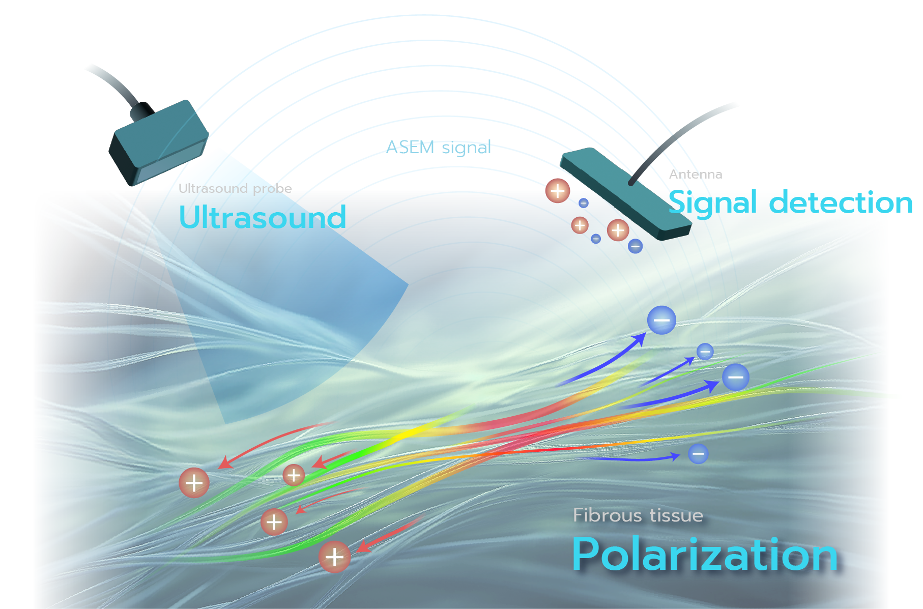
We discovered a phenomenon in which electricity and magnetism are induced inside biological tissues or materials by irradiating ultrasound.
The electromagnetic signals (Acousto-Electromagnetic Signals: ASEM signals) generated in this process are the basis for a sensing technology we are developing into a practical application.This “ASEM method” is based on fundamental research conducted by the Ikushima Laboratory, Tokyo University of Agriculture and Technology.
ref.)
K. Ikushima, Progress Review, Jpn. J. Appl. Phys. 62, SJ0802 (2023)
When force is applied to a material, distortion may cause it to generate electricity.
This phenomenon is known as the piezoelectric effect, and it is commonly observed in crystals and certain ceramics, being widely used in piezoelectric devices such as mobile speakers and sonar systems.
Originally, the piezoelectric effect is a property observed in inorganic single-crystal materials, and its characteristics are determined by the order and symmetry of atomic arrangements in the crystal.
We confirmed that even in living biological tissues, the piezoelectric effect appears in structures with ordered molecular arrangements, such as collagen fibers (crystallinity and orientation).
By capturing the electricity generated by ultrasound (sound pressure) with an externally placed receiving antenna, we successfully achieved non-invasive detection and imaging of biological piezoelectric signals.
This demonstrates that measuring biological piezoelectric signals enables quantitative assessment of fiber orientation and collagen accumulation, providing insight into pathological tissue conditions.
ref.)
Progress Review, “Acoustically induced electric and magnetic polarizations and their sensing applications”, Jpn. J. Appl. Phys. 62, SJ0802 (2023)


Non-Invasive Bio-Sensing
- Fibrosis diagnosis in cancer/chronic disease
- Healing assessment of fractures/tendon injuries
- Exercise effect visualization & quantification
In recent years, ultrasound imaging devices have been gaining attention as tools that enable rapid bedside examinations in outpatient and home care settings, with growing demand for their role in improving the quality of medical care and patients’ quality of life (QOL).
In addition to conventional “visual examinations” and “observation of internal structures via echo”, the ASEM method offers a new perspective closer to pathological evaluation (= a “third eye”), enabling more precise diagnoses and faster treatment decisions.
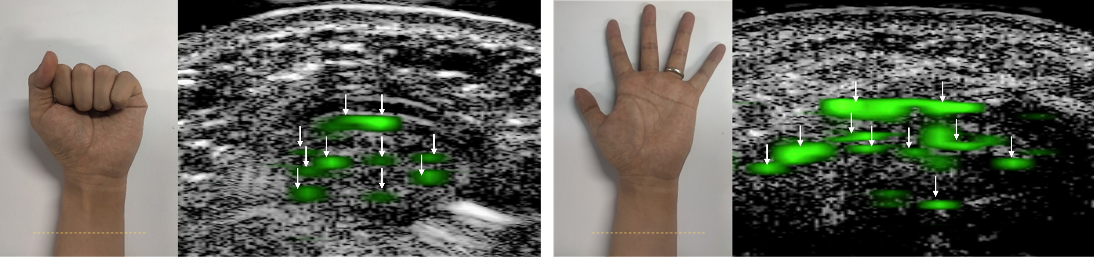
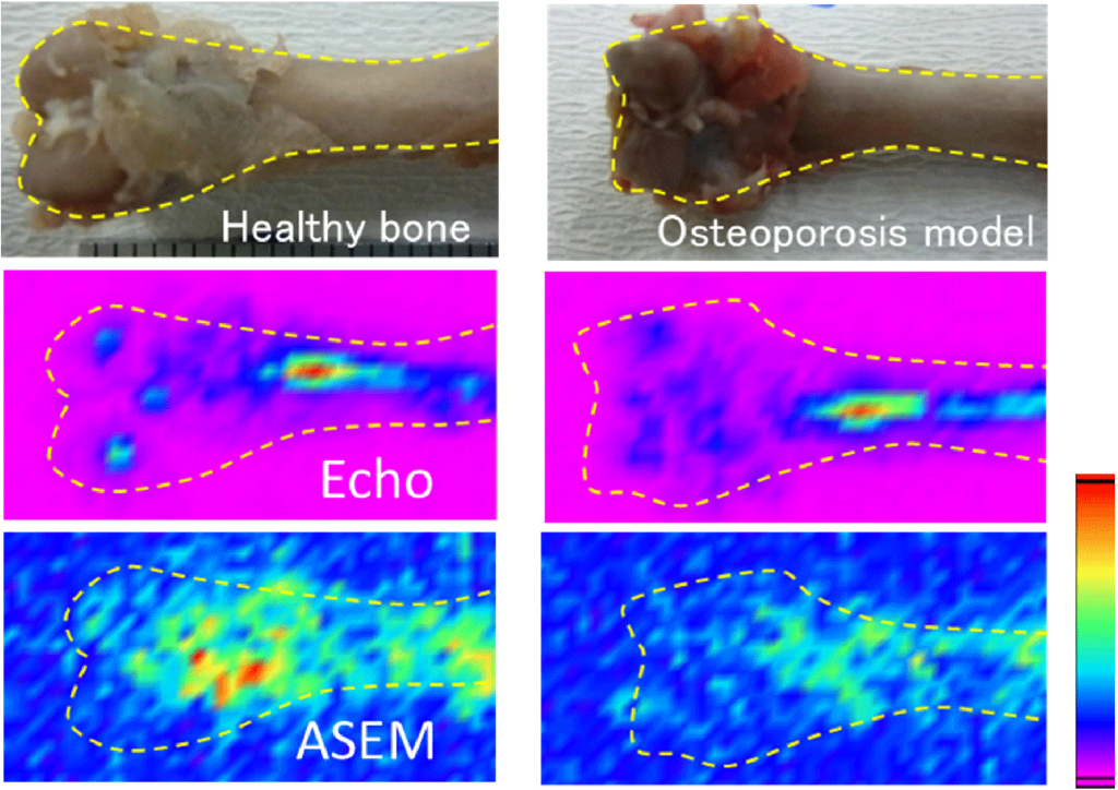
ASEM is a technique that visualizes electrical signals (ASEM signals) induced by ultrasound pressure in fibrous biological tissues such as bones, tendons, ligaments, and arterial walls or valves.
From these signals, it is possible to image the “quality” of tissues—such as the crystallinity and orientation of collagen and elastin.
In particular, it is being applied to the diagnosis of organ fibrosis in cancer and chronic diseases, and to the quantification and visualization of treatment effects for fractures and tendon ruptures, as well as rehabilitation outcomes.
Collagen is a key structural protein in our bodies, not only forming the musculoskeletal system but also being widely distributed across organs and blood vessels.
Fractures and joint diseases are a leading cause of mobility issues and the need for nursing care and support, and as global aging accelerates, maintaining physical function is critical for independent living.
Early recovery from fractures or tendon injuries is crucial for both individual QOL and social reintegration.
This project aims to expand applications beyond clinical diagnosis and treatment, to include rehabilitation and daily health management.
By utilizing ASEM, we hope to help people better understand their physical condition and enjoy appropriate exercise, enabling them to live healthy and fulfilling lives.
ref.)
Nature Research Highlights “ The body electric: soft tissue makes electricity under stress“
APS Physics: Soft Biological Tissues Can Be Piezoelectric.
“Electric Polarization of Soft Biological Tissues Induced by Ultrasound Waves”, Phys. Rev. Lett.123, 238101 (2019) (Original manuscript, Supplemental material)
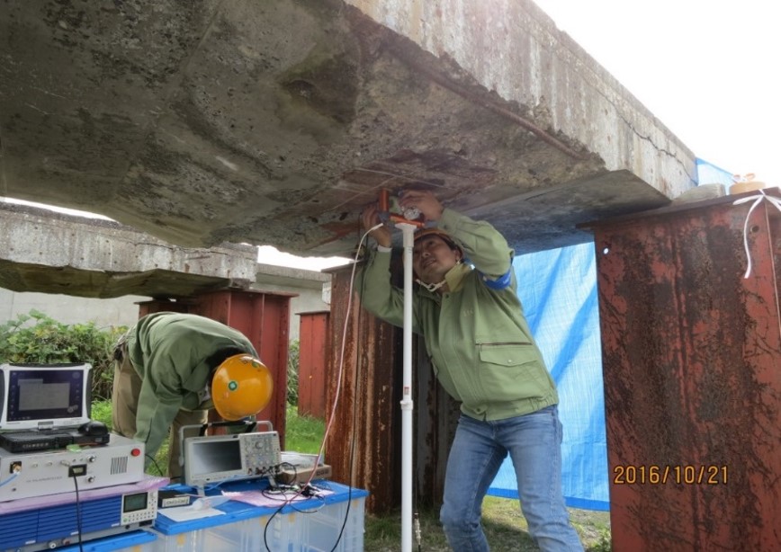
Non-Destructive Material Sensing
- Steel defect inspection
- Corrosion & degradation testing
- Residual stress assessment
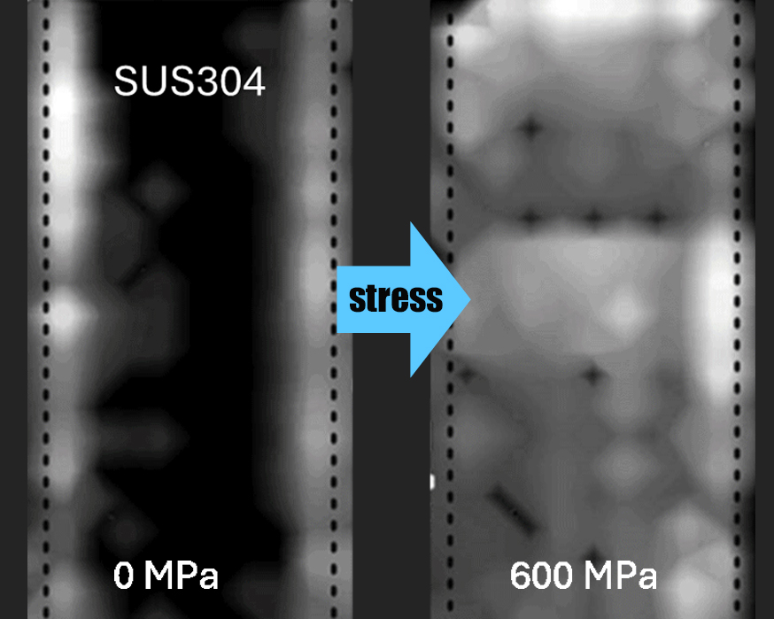
The magnetic properties of steel—such as crystal phase, grain size, residual stress, corrosion, defects, and heat treatment history—are sensitive to various factors, making them effective indicators for non-destructive testing.
Our advanced localized magnetic measurement system, unlike conventional magnetic inspection methods, enables non-contact or semi-contact imaging and quantification of subtle magnetic variations using ultrasound.
As a non-destructive testing solution, it allows accurate assessment of steel conditions, improving safety and durability, and contributing to reduced production costs.
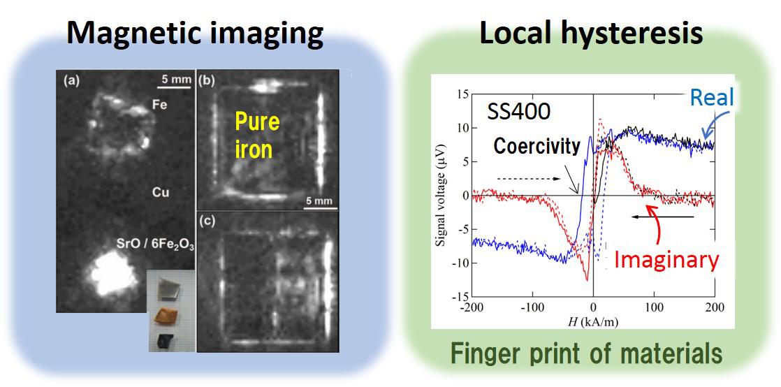
In ferromagnetic materials like steel, ultrasound generates magnetic signals (ASEM signals). These signals reflect the internal magnetic structure and state. By reading them, it’s possible to visualize the internal conditions of the material non-destructively.
ASEM enables magnetic imaging and hysteresis measurement using ultrasound.
It detects signs of corrosion, degradation, embrittlement, residual stress, and metal fatigue, as well as subtle material inhomogeneities invisible to the eye.
This technology shows promise for non-destructive inspection of materials like steel.

Technology Timeline
Evolution of ASEM
2006
2013
2015
2018
2019
2022
2023
2023 Nov.

Electromagnetic signals are generated From bone and wood by ultrasound
Magnetic imaging by ultrasound
Magnetic hysteresis measurements by ultrasound
Flaw detection for thin steel
Piezoelectric imaging of bone
Piezoelectricity of soft biological tissues
Nature Research Highlights
NDE of residual stress in steel
ASEM tomogram of human upper limb
ASEMtech Inc. established